Copyright
©The Author(s) 2017.
World J Clin Cases. Feb 16, 2017; 5(2): 61-66
Published online Feb 16, 2017. doi: 10.12998/wjcc.v5.i2.61
Published online Feb 16, 2017. doi: 10.12998/wjcc.v5.i2.61
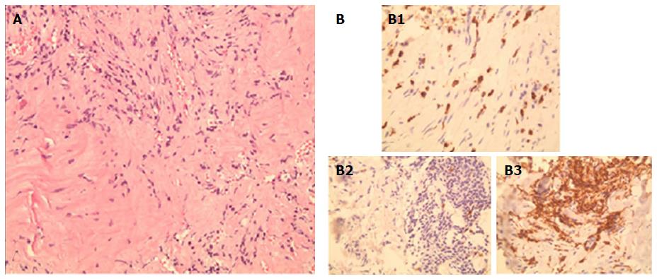
Figure 3 (A) Heavy infiltration of lymphocytes, plasma cells, and histiocytes, within a background of spindle-shaped fibroblasts and myofibroblasts, arrayed in fascicles (B).
Immunostaining of tissues showing positivity for CD3 (B1), negativity for CD20 (B2), and positivity for CD138 (B3), respectively.
- Citation: Degheili JA, Kanj NA, Koubaissi SA, Nasser MJ. Indolent lung opacity: Ten years follow-up of pulmonary inflammatory pseudo-tumor. World J Clin Cases 2017; 5(2): 61-66
- URL: https://www.wjgnet.com/2307-8960/full/v5/i2/61.htm
- DOI: https://dx.doi.org/10.12998/wjcc.v5.i2.61


