Copyright
©The Author(s) 2017.
World J Clin Cases. Feb 16, 2017; 5(2): 61-66
Published online Feb 16, 2017. doi: 10.12998/wjcc.v5.i2.61
Published online Feb 16, 2017. doi: 10.12998/wjcc.v5.i2.61
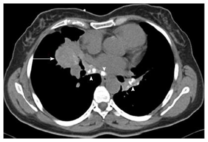
Figure 2 Computed tomography of the chest revealing a right middle lobe mass (arrow), with multiple calcified hilar and mediastinal lymph nodes (arrow heads).
- Citation: Degheili JA, Kanj NA, Koubaissi SA, Nasser MJ. Indolent lung opacity: Ten years follow-up of pulmonary inflammatory pseudo-tumor. World J Clin Cases 2017; 5(2): 61-66
- URL: https://www.wjgnet.com/2307-8960/full/v5/i2/61.htm
- DOI: https://dx.doi.org/10.12998/wjcc.v5.i2.61


