Copyright
©The Author(s) 2017.
World J Clin Cases. Dec 16, 2017; 5(12): 446-452
Published online Dec 16, 2017. doi: 10.12998/wjcc.v5.i12.446
Published online Dec 16, 2017. doi: 10.12998/wjcc.v5.i12.446
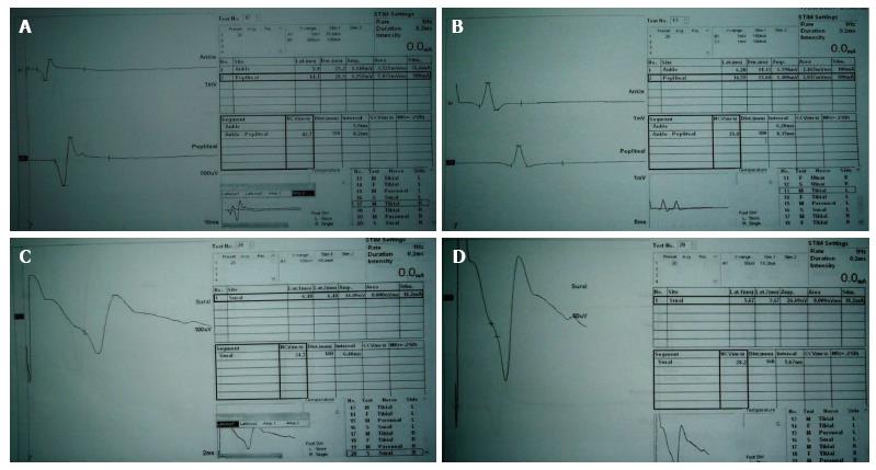
Figure 1 Nerve conduction velocity study traces of the right (A) and left tibial (B) nerves and right (C) and left sural (D) nerves show prolonged distal latencies, reduced motor and sensory conduction velocities and reduced motor and sensory action potentials (amplitudes).
- Citation: Hamed SA. Topiramate induced peripheral neuropathy: A case report and review of literature. World J Clin Cases 2017; 5(12): 446-452
- URL: https://www.wjgnet.com/2307-8960/full/v5/i12/446.htm
- DOI: https://dx.doi.org/10.12998/wjcc.v5.i12.446


