Copyright
©The Author(s) 2017.
World J Clin Cases. Dec 16, 2017; 5(12): 440-445
Published online Dec 16, 2017. doi: 10.12998/wjcc.v5.i12.440
Published online Dec 16, 2017. doi: 10.12998/wjcc.v5.i12.440
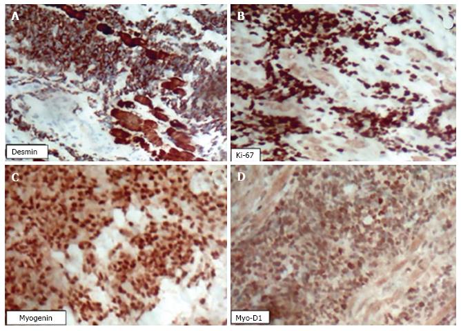
Figure 4 An immunohistochemical analysis was performed on a biopsied tumour fragment from the left maxillary sinus.
A: Immunohistochemical analysis showed positiveness to anti-Desmin antibody with dual cytoplasmatic and nuclear staining; B: The same pattern was observed against anti-Ki67 (B) showing intense positiveness and high rate of cell proliferation; C and D: Anti-myogenin and MYO-D1 were positively found on nuclear staining leading to RMS lineage supposition.
- Citation: de Melo ACR, Lyra TC, Ribeiro ILA, da Paz AR, Bonan PRF, de Castro RD, Valença AMG. Embryonal rhabdomyosarcoma in the maxillary sinus with orbital involvement in a pediatric patient: Case report. World J Clin Cases 2017; 5(12): 440-445
- URL: https://www.wjgnet.com/2307-8960/full/v5/i12/440.htm
- DOI: https://dx.doi.org/10.12998/wjcc.v5.i12.440


