Copyright
©The Author(s) 2017.
World Journal of Clinical Cases. Nov 16, 2017; 5(11): 397-402
Published online Nov 16, 2017. doi: 10.12998/wjcc.v5.i11.397
Published online Nov 16, 2017. doi: 10.12998/wjcc.v5.i11.397
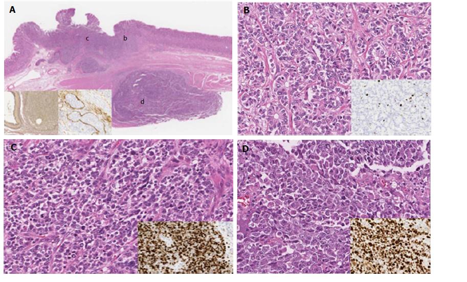
Figure 1 Histological findings of an HE-stained section of a resected specimen.
A: Low-magnification image of the surgical specimen. The tumor component was composed of two lesions (a submucosal lesion and an intramucosal lesion). These two lesions were separated by muscle layer (inset: left, EVG staining; right, D2-40 immunohistochemical staining); B: NET G2 component (inset: Ki-67 positive rate, 6.5%); C: NET G3 component (inset: Ki-67 positive rate, 99.5%); D: Large cell NEC component (inset: Ki-67 positive rate, 88.1%). NEC: Neuroendocrine carcinoma; NET: Neuroendocrine tumor.
- Citation: Uesugi N, Sugimoto R, Eizuka M, Fujita Y, Osakabe M, Koeda K, Kosaka T, Yanai S, Ishida K, Sasaki A, Matsumoto T, Sugai T. Case of gastric neuroendocrine carcinoma showing an interesting tumorigenic pathway. World Journal of Clinical Cases 2017; 5(11): 397-402
- URL: https://www.wjgnet.com/2307-8960/full/v5/i11/397.htm
- DOI: https://dx.doi.org/10.12998/wjcc.v5.i11.397


