Copyright
©The Author(s) 2017.
World J Clin Cases. Oct 16, 2017; 5(10): 373-377
Published online Oct 16, 2017. doi: 10.12998/wjcc.v5.i10.373
Published online Oct 16, 2017. doi: 10.12998/wjcc.v5.i10.373
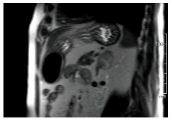
Figure 2 Coronal magnetic resonance imaging of left adrenal ganglioneuroma: A coronal T1-weighted out-of-phase image shows intracellular lipid and no signal loss within the lesion.
- Citation: Mylonas KS, Schizas D, Economopoulos KP. Adrenal ganglioneuroma: What you need to know. World J Clin Cases 2017; 5(10): 373-377
- URL: https://www.wjgnet.com/2307-8960/full/v5/i10/373.htm
- DOI: https://dx.doi.org/10.12998/wjcc.v5.i10.373


