Copyright
©The Author(s) 2016.
World J Clinical Cases. Sep 16, 2016; 4(9): 302-305
Published online Sep 16, 2016. doi: 10.12998/wjcc.v4.i9.302
Published online Sep 16, 2016. doi: 10.12998/wjcc.v4.i9.302
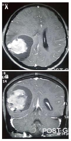
Figure 1 Post contrast magnetic resonance imaging axial (A) and coronal (B) images shows large heterogeneous lesion in right parieto-occipital region with effacement of ipsilateral lateral ventricle.
- Citation: Meena US, Sharma S, Chopra S, Jain SK. Gliosarcoma: A rare variant of glioblastoma multiforme in paediatric patient: Case report and review of literature. World J Clinical Cases 2016; 4(9): 302-305
- URL: https://www.wjgnet.com/2307-8960/full/v4/i9/302.htm
- DOI: https://dx.doi.org/10.12998/wjcc.v4.i9.302


