Copyright
©The Author(s) 2016.
World J Clinical Cases. Sep 16, 2016; 4(9): 290-295
Published online Sep 16, 2016. doi: 10.12998/wjcc.v4.i9.290
Published online Sep 16, 2016. doi: 10.12998/wjcc.v4.i9.290
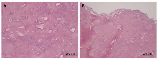
Figure 2 Histological analysis of the lesion.
A: Central region of the lesion, where a cementoid structure with blood vessels was observed, presenting superimposed lamellae and basophilic material; B: Peripheral region of the lesion that presented irregular fibrous tissue, with tissue having a cementoid aspect and presence of blood vessels (HE-50 × magnification).
- Citation: Costa BC, de Oliveira GJPL, Chaves MDGAM, da Costa RR, Gabrielli MFR, Guerreiro-Tanomaru JM, Tanomaru-Filho M. Surgical treatment of cementoblastoma associated with apicoectomy and endodontic therapy: Case report. World J Clinical Cases 2016; 4(9): 290-295
- URL: https://www.wjgnet.com/2307-8960/full/v4/i9/290.htm
- DOI: https://dx.doi.org/10.12998/wjcc.v4.i9.290


