Copyright
©The Author(s) 2016.
World J Clinical Cases. Sep 16, 2016; 4(9): 281-284
Published online Sep 16, 2016. doi: 10.12998/wjcc.v4.i9.281
Published online Sep 16, 2016. doi: 10.12998/wjcc.v4.i9.281
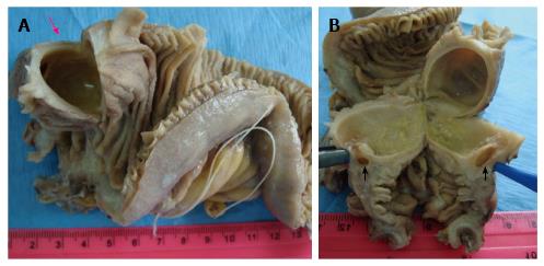
Figure 1 Macroscopic findings of the jejunal spherical duplication cyst.
The submucosal cystic tumor is covered by intact jejunal mucosa and is filled with clear fluid (pink arrow, A). Its wall shares the muscularis layer of the adjacent jejunum and a communication with the intestinal lumen is seen (black arrows, B).
- Citation: Gurzu S, Bara Jr T, Bara T, Fetyko A, Jung I. Cystic jejunal duplication with Heinrich’s type I ectopic pancreas, incidentally discovered in a patient with pancreatic tail neoplasm. World J Clinical Cases 2016; 4(9): 281-284
- URL: https://www.wjgnet.com/2307-8960/full/v4/i9/281.htm
- DOI: https://dx.doi.org/10.12998/wjcc.v4.i9.281


