Copyright
©The Author(s) 2016.
World J Clin Cases. Aug 16, 2016; 4(8): 248-252
Published online Aug 16, 2016. doi: 10.12998/wjcc.v4.i8.248
Published online Aug 16, 2016. doi: 10.12998/wjcc.v4.i8.248
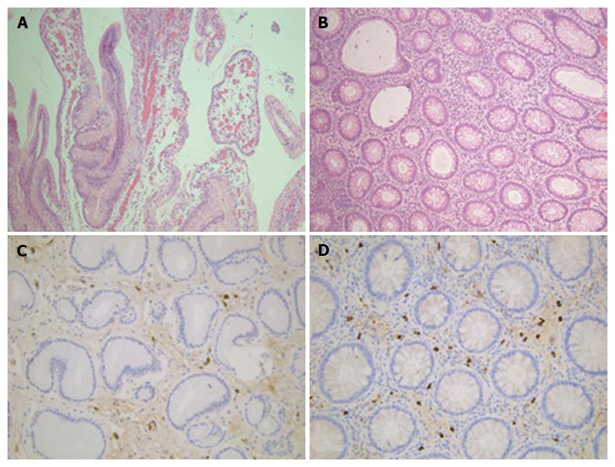
Figure 3 Histopathologic examination of biopsies.
A: Histopathology examination of gastric polyp showing inflammatory and hyperplasia change (hematoxylin-eosin stain, × 20); B: Histopathology examination of colonic polyp showing inflammatory change (hematoxylin-eosin stain, × 10); C: IgG4 mononuclear cell staining in gastric polyp (× 20); D: IgG4 mononuclear cell staining in gastric polyp (hematoxylin-eosin stain, × 20).
- Citation: Fan RY, Wang XW, Xue LJ, An R, Sheng JQ. Cronkhite-Canada syndrome polyps infiltrated with IgG4-positive plasma cells. World J Clin Cases 2016; 4(8): 248-252
- URL: https://www.wjgnet.com/2307-8960/full/v4/i8/248.htm
- DOI: https://dx.doi.org/10.12998/wjcc.v4.i8.248


