Copyright
©The Author(s) 2016.
World J Clin Cases. Aug 16, 2016; 4(8): 238-242
Published online Aug 16, 2016. doi: 10.12998/wjcc.v4.i8.238
Published online Aug 16, 2016. doi: 10.12998/wjcc.v4.i8.238
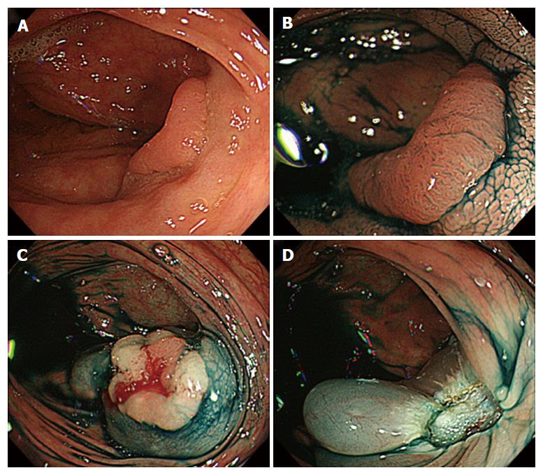
Figure 1 Colonoscopy images when endoscopic mucosal resection was performed.
A: An 18-mm slightly reddish elevated lesion was recognized on the transverse colon; B: Type 3L pit pattern was recognized with indigo was recognized; C: Endoscopic mucosal resection (EMR) was performed; D: There were no apparent findings that suggested perforation at the ulcer floor immediately after EMR.
- Citation: Inoki K, Sakamoto T, Sekiguchi M, Yamada M, Nakajima T, Matsuda T, Saito Y. Successful endoscopic closure of a colonic perforation one day after endoscopic mucosal resection of a lesion in the transverse colon. World J Clin Cases 2016; 4(8): 238-242
- URL: https://www.wjgnet.com/2307-8960/full/v4/i8/238.htm
- DOI: https://dx.doi.org/10.12998/wjcc.v4.i8.238


