Copyright
©The Author(s) 2016.
World J Clin Cases. Jul 16, 2016; 4(7): 172-176
Published online Jul 16, 2016. doi: 10.12998/wjcc.v4.i7.172
Published online Jul 16, 2016. doi: 10.12998/wjcc.v4.i7.172
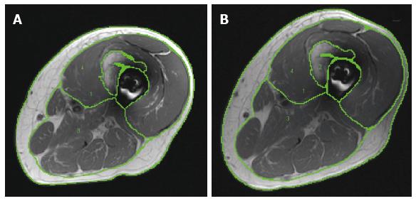
Figure 1 Representative magnetic resonance images of (A) pre- and (B) post-intervention showing different regions of interest (1) whole thigh skeletal muscle cross-sectional areas; (2) Heterotopic ossification bone formation; (3) posterior and medial compartments mid-thigh muscles; (4) knee extensor cross-sectional area.
- Citation: Moore PD, Gorgey AS, Wade RC, Khalil RE, Lavis TD, Khan R, Adler RA. Neuromuscular electrical stimulation and testosterone did not influence heterotopic ossification size after spinal cord injury: A case series. World J Clin Cases 2016; 4(7): 172-176
- URL: https://www.wjgnet.com/2307-8960/full/v4/i7/172.htm
- DOI: https://dx.doi.org/10.12998/wjcc.v4.i7.172


