Copyright
©The Author(s) 2016.
World J Clin Cases. May 16, 2016; 4(5): 127-129
Published online May 16, 2016. doi: 10.12998/wjcc.v4.i5.127
Published online May 16, 2016. doi: 10.12998/wjcc.v4.i5.127
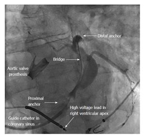
Figure 1 Balloon occlusion venogram in the coronary sinus.
The proximal and distal anchors of the annuloplasty device are shown within the coronary sinus, which is filled with contrast to show target branches for the LV lead. LV: Left ventricular.
- Citation: Swampillai J. Cardiac resynchronisation therapy after percutaneous mitral annuloplasty. World J Clin Cases 2016; 4(5): 127-129
- URL: https://www.wjgnet.com/2307-8960/full/v4/i5/127.htm
- DOI: https://dx.doi.org/10.12998/wjcc.v4.i5.127


