Copyright
©The Author(s) 2016.
World J Clin Cases. Feb 16, 2016; 4(2): 38-48
Published online Feb 16, 2016. doi: 10.12998/wjcc.v4.i2.38
Published online Feb 16, 2016. doi: 10.12998/wjcc.v4.i2.38
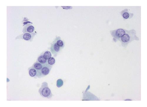
Figure 3 A cellular specimen composed of Hürthle cells arranged in loosely cohesive sheets or isolated in a case diagnosed as Hürthle cell adenoma (× 40 pap stain on ThinPrep slide) (diagnostic categories IV).
- Citation: Misiakos EP, Margari N, Meristoudis C, Machairas N, Schizas D, Petropoulos K, Spathis A, Karakitsos P, Machairas A. Cytopathologic diagnosis of fine needle aspiration biopsies of thyroid nodules. World J Clin Cases 2016; 4(2): 38-48
- URL: https://www.wjgnet.com/2307-8960/full/v4/i2/38.htm
- DOI: https://dx.doi.org/10.12998/wjcc.v4.i2.38


