Copyright
©The Author(s) 2016.
World J Clin Cases. Dec 16, 2016; 4(12): 419-422
Published online Dec 16, 2016. doi: 10.12998/wjcc.v4.i12.419
Published online Dec 16, 2016. doi: 10.12998/wjcc.v4.i12.419
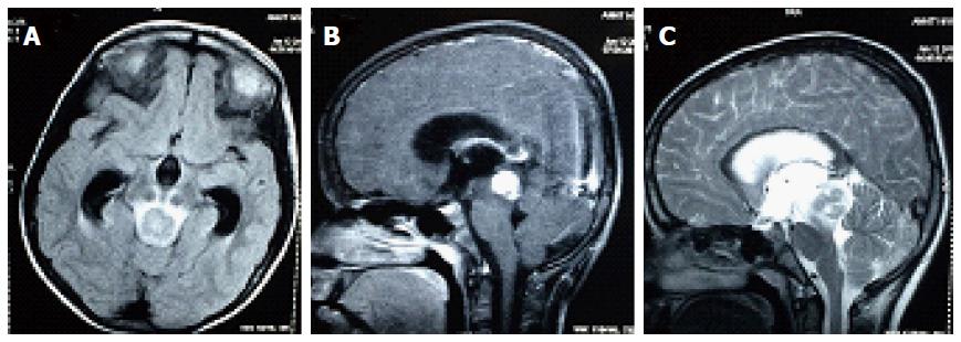
Figure 1 Axial and sagittal section of contrast enhanced magnetic resonance imaging.
It shows a lesion 1.7 cm × 1.6 cm × 1.6 cm in the mid brain and tectum with intense contrast enhancement. The lesion was mildly hypo-intense in T1 and heterogeneously hyper-intense on T2W image with few hypo-intense areas. There edema was extending to mid part of pons.
- Citation: Benson R, Mallick S, Purkait S, Suri V, Haresh KP, Gupta S, Sharma D, Julka PK, Rath GK. Primary pediatric mid-brain lymphoma: Report of a rare pediatric tumor in a rare location. World J Clin Cases 2016; 4(12): 419-422
- URL: https://www.wjgnet.com/2307-8960/full/v4/i12/419.htm
- DOI: https://dx.doi.org/10.12998/wjcc.v4.i12.419


