Copyright
©The Author(s) 2016.
World J Clin Cases. Dec 16, 2016; 4(12): 413-418
Published online Dec 16, 2016. doi: 10.12998/wjcc.v4.i12.413
Published online Dec 16, 2016. doi: 10.12998/wjcc.v4.i12.413
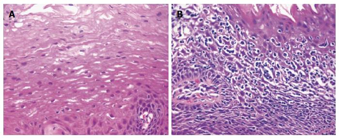
Figure 2 Histopathologic findings.
A: Normal esophageal mucosa with stratified squamous epithelium and no inflammatory cells present; B: Lymphocytic esophagitis with marked spongiosis and intraepithelial lymphocyte infiltration in basal and peripapillary fields. No granulocytes present (reproduced from ref. [3]).
- Citation: Jideh B, Keegan A, Weltman M. Lymphocytic esophagitis: Report of three cases and review of the literature. World J Clin Cases 2016; 4(12): 413-418
- URL: https://www.wjgnet.com/2307-8960/full/v4/i12/413.htm
- DOI: https://dx.doi.org/10.12998/wjcc.v4.i12.413


