Copyright
©The Author(s) 2016.
World J Clin Cases. Nov 16, 2016; 4(11): 380-384
Published online Nov 16, 2016. doi: 10.12998/wjcc.v4.i11.380
Published online Nov 16, 2016. doi: 10.12998/wjcc.v4.i11.380
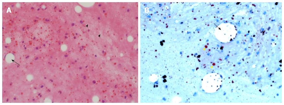
Figure 4 Histopathology (A) and Oil red O stain (B) of a cell block of the left pleural fluid.
A: Macrophages containing large fat vacuoles (black arrow) and globules of fat in the background (arrow heads). Macrophages show eccentric nuclei and “Empty looking” cytoplasm. Most of the fat has been dissolved and removed during processing, having been dissolved by the xylene and alcohol. A few mixed inflammatory cells are seen in the background (Haematoxylin and eosin, × 40); B: An Oil red O stain demonstrates fat globules of varying sizes stained orange (yellow arrow heads). Some globules are still present in the macrophages (black arrow), but most of them are extracellular, within the chylous fluid (Oil red O stain × 40).
- Citation: Idris K, Sebastian M, Hefny AF, Khan NH, Abu-Zidan FM. Blunt traumatic tension chylothorax: Case report and mini-review of the literature. World J Clin Cases 2016; 4(11): 380-384
- URL: https://www.wjgnet.com/2307-8960/full/v4/i11/380.htm
- DOI: https://dx.doi.org/10.12998/wjcc.v4.i11.380


