Copyright
©The Author(s) 2016.
World J Clin Cases. Nov 16, 2016; 4(11): 380-384
Published online Nov 16, 2016. doi: 10.12998/wjcc.v4.i11.380
Published online Nov 16, 2016. doi: 10.12998/wjcc.v4.i11.380
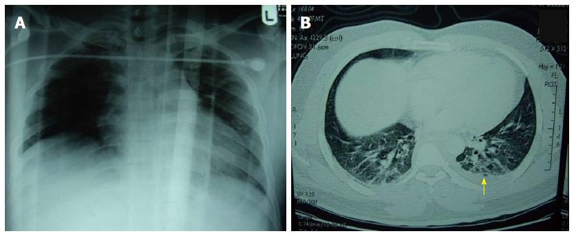
Figure 1 Antero-posterior chest X-ray performed at presentation (A) and contrast enhanced computed tomography (B).
A: Fracture right clavicle and mild haziness of the left lung field; B: Mild pleural effusion of the left side (yellow arrow).
- Citation: Idris K, Sebastian M, Hefny AF, Khan NH, Abu-Zidan FM. Blunt traumatic tension chylothorax: Case report and mini-review of the literature. World J Clin Cases 2016; 4(11): 380-384
- URL: https://www.wjgnet.com/2307-8960/full/v4/i11/380.htm
- DOI: https://dx.doi.org/10.12998/wjcc.v4.i11.380


