Copyright
©The Author(s) 2016.
World J Clin Cases. Nov 16, 2016; 4(11): 364-368
Published online Nov 16, 2016. doi: 10.12998/wjcc.v4.i11.364
Published online Nov 16, 2016. doi: 10.12998/wjcc.v4.i11.364
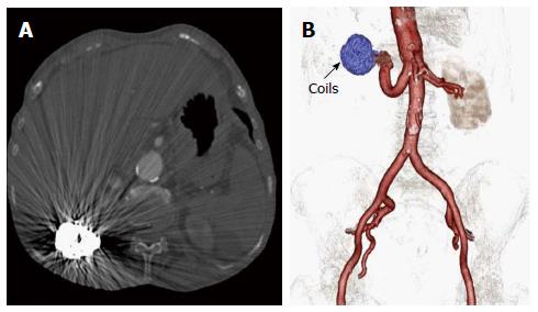
Figure 3 Images reveal resolution of ascites and occlusion of the renal arteriovenous fistula.
A: Axial CT image following successful coil occlusion of fistula reveal interval lack of opacification of right renal venous structures and resolution of ascites. Note beam hardening related streak artifacts from the embolization coils within the right kidney; B: 3D Volume rendered image using post-processing segmentation tools clearly reveals the embolization coils and non-visualization of IVC and renal venous system in this arterial phase scan suggesting occlusion of the renal arteriovenous fistula. Note residual ectatic right renal artery. CT: Computed tomography; IVC: Inferior vena cava.
- Citation: Nagpal P, Bathla G, Saboo SS, Khandelwal A, Goyal A, Rybicki FJ, Steigner ML. Giant idiopathic renal arteriovenous fistula managed by coils and amplatzer device: Case report and literature review. World J Clin Cases 2016; 4(11): 364-368
- URL: https://www.wjgnet.com/2307-8960/full/v4/i11/364.htm
- DOI: https://dx.doi.org/10.12998/wjcc.v4.i11.364


