Copyright
©The Author(s) 2016.
World J Clin Cases. Oct 16, 2016; 4(10): 344-350
Published online Oct 16, 2016. doi: 10.12998/wjcc.v4.i10.344
Published online Oct 16, 2016. doi: 10.12998/wjcc.v4.i10.344
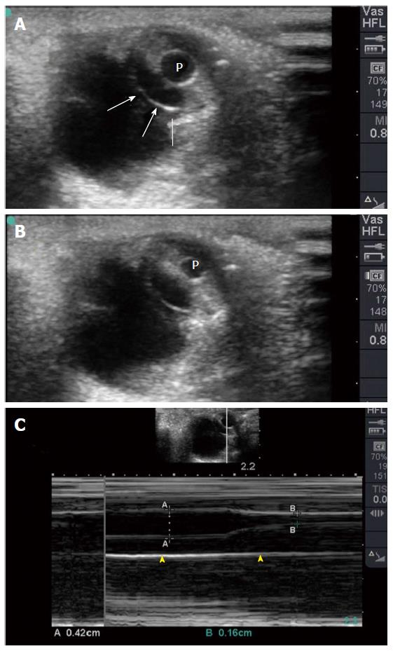
Figure 11 Coronal eye view (A) showing the pupil and edge of the iris (white arrows).
The pupil (P) constricts when light is applied to the closed eye (B). M Mode (C) accurately measures the size of the pupil which constricted from 4.2 mm to 1.6 mm to light reflex.
- Citation: Abu-Zidan FM, Balac K, Bhatia CA. Surgeon-performed point-of-care ultrasound in severe eye trauma: Report of two cases. World J Clin Cases 2016; 4(10): 344-350
- URL: https://www.wjgnet.com/2307-8960/full/v4/i10/344.htm
- DOI: https://dx.doi.org/10.12998/wjcc.v4.i10.344


