Copyright
©The Author(s) 2016.
World J Clin Cases. Oct 16, 2016; 4(10): 344-350
Published online Oct 16, 2016. doi: 10.12998/wjcc.v4.i10.344
Published online Oct 16, 2016. doi: 10.12998/wjcc.v4.i10.344
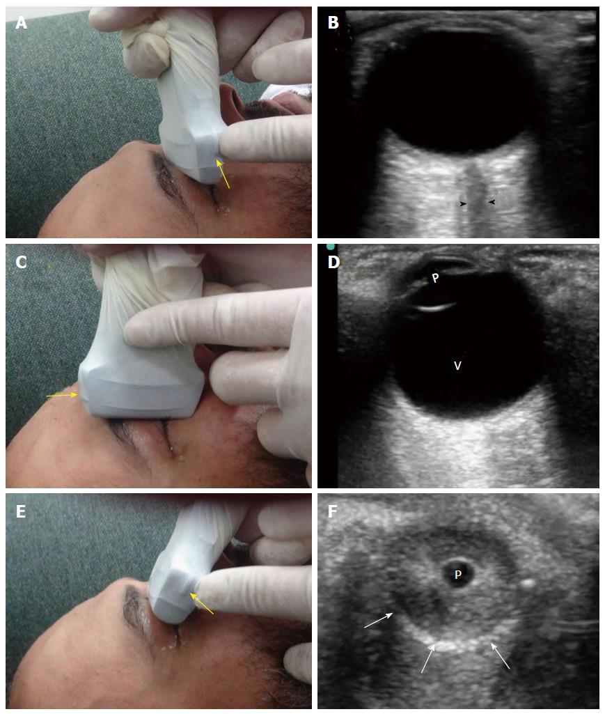
Figure 10 There are three common views to examine the eye.
The transverse antero-posterior view (A) is useful for examining the optic nerve (B, arrow heads). The sagittal antero-posterior view (C) is useful in visualizing the anterior and posterior chambers of the eye (D, V: Vitreous; p: Pupil). The coronal view (E) is useful for examining the pupil (F, p: Pupil, white arrows: Edge of the iris). The marker of the probe (yellow arrows) should point to the right side of the patient or upwards.
- Citation: Abu-Zidan FM, Balac K, Bhatia CA. Surgeon-performed point-of-care ultrasound in severe eye trauma: Report of two cases. World J Clin Cases 2016; 4(10): 344-350
- URL: https://www.wjgnet.com/2307-8960/full/v4/i10/344.htm
- DOI: https://dx.doi.org/10.12998/wjcc.v4.i10.344


