Copyright
©The Author(s) 2016.
World J Clin Cases. Oct 16, 2016; 4(10): 328-332
Published online Oct 16, 2016. doi: 10.12998/wjcc.v4.i10.328
Published online Oct 16, 2016. doi: 10.12998/wjcc.v4.i10.328
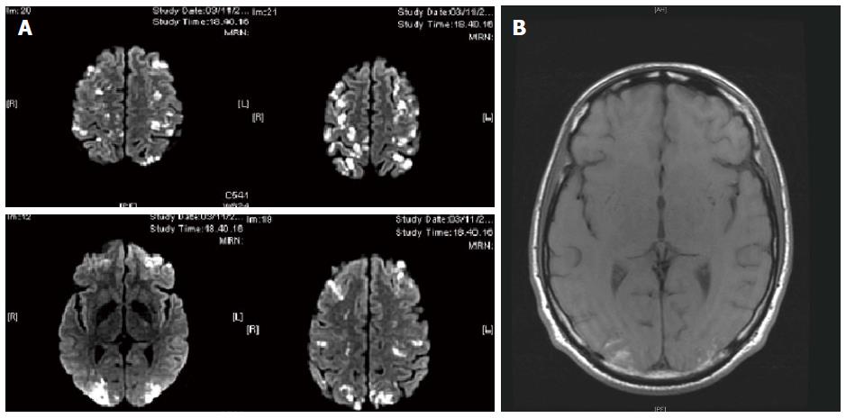
Figure 2 Brain magnetic resonance imaging showed multiple small focal lesions, with the most serious in the occipital regions and others in the frontal areas.
A: Diffusion weighted imaging was consistent with ischemic multiple lesions; B: Spontaneous hyperintensity in T1, indicating blood suffusions and suggestive of vasculitis. Following this magnetic resonance imaging, the patient performed a campimetric exam that showed an asymmetric bilateral visual loss, which did not ever completely resolve despite therapy.
- Citation: Fraticelli P, Kafyeke A, Mattioli M, Martino GP, Murri M, Gabrielli A. Idiopathic hypereosinophilic syndrome presenting with severe vasculitis successfully treated with imatinib. World J Clin Cases 2016; 4(10): 328-332
- URL: https://www.wjgnet.com/2307-8960/full/v4/i10/328.htm
- DOI: https://dx.doi.org/10.12998/wjcc.v4.i10.328


