Copyright
©The Author(s) 2016.
World J Clin Cases. Oct 16, 2016; 4(10): 328-332
Published online Oct 16, 2016. doi: 10.12998/wjcc.v4.i10.328
Published online Oct 16, 2016. doi: 10.12998/wjcc.v4.i10.328
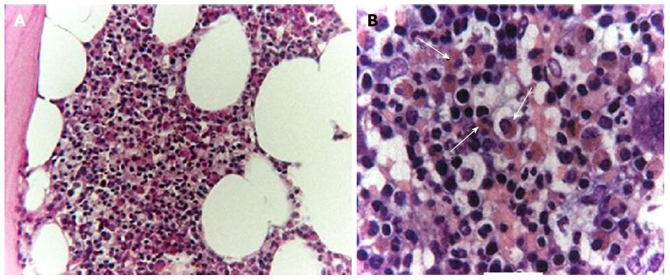
Figure 1 Bone marrow biopsy.
A: Hematoxylin and eosin (H and E)-stained section of paraffin-embedded bone marrow showing expansion of the myeloid cellular line with marked increase in granulated eosinophilic cells (100 ×); B: High-power view of the diffuse eosinophilic infiltration (white arrows), without any detectable morphological signs of abnormal proliferation (400 ×).
- Citation: Fraticelli P, Kafyeke A, Mattioli M, Martino GP, Murri M, Gabrielli A. Idiopathic hypereosinophilic syndrome presenting with severe vasculitis successfully treated with imatinib. World J Clin Cases 2016; 4(10): 328-332
- URL: https://www.wjgnet.com/2307-8960/full/v4/i10/328.htm
- DOI: https://dx.doi.org/10.12998/wjcc.v4.i10.328


