Copyright
©The Author(s) 2015.
World J Clin Cases. Sep 16, 2015; 3(9): 779-788
Published online Sep 16, 2015. doi: 10.12998/wjcc.v3.i9.779
Published online Sep 16, 2015. doi: 10.12998/wjcc.v3.i9.779
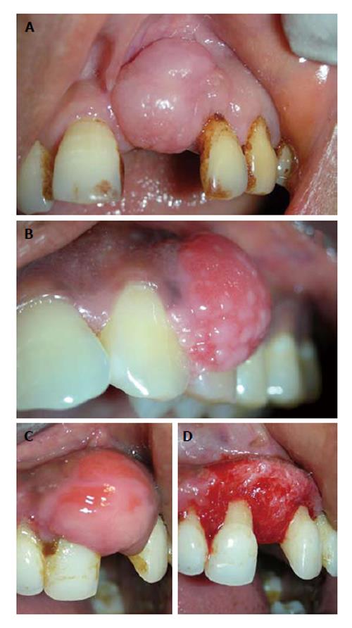
Figure 1 Fibrous epulis and its subtypes.
A: Peripheral fibroma, presenting as pink firm, uninflammed mass growing from under the gingiva; B: Peripheral cementifying fibroma, a subcategory of fibroma, shows additional foci of cementicles; C and D: Surgical exposure of the lesion showing extensive bone formation in the core of the lesion. Also presence of bony trabeculae was seen histologically.
- Citation: Agrawal AA. Gingival enlargements: Differential diagnosis and review of literature. World J Clin Cases 2015; 3(9): 779-788
- URL: https://www.wjgnet.com/2307-8960/full/v3/i9/779.htm
- DOI: https://dx.doi.org/10.12998/wjcc.v3.i9.779


