Copyright
©The Author(s) 2015.
World J Clin Cases. Sep 16, 2015; 3(9): 765-773
Published online Sep 16, 2015. doi: 10.12998/wjcc.v3.i9.765
Published online Sep 16, 2015. doi: 10.12998/wjcc.v3.i9.765
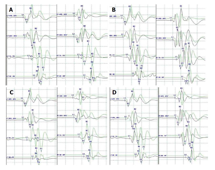
Figure 2 Representative case demonstrating clinical usefulness of intraoperative neuromonitoring in spinal surgery.
A: MEP after applying rod to the screw heads using derotation maneuver and cantilever maneuver. The amplitude of MEP (black line) at both lower extremities decreased more than 50% compared with the baseline amplitude (green line); B: The amplitude of MEP recovered after correction release by removal of the rods and set screws; C: The amplitude of MEP re-deteriorated after reassembly of the implants; D: The amplitude of MEP recovered finally after raising MAP and administration of dexamethasone. APB: Abductor pollicis brevis; ADQ: Abductor digiti quinti; TA: Tibialis anterior; AH: Abductor halluces; MEP: Motor evoked potential; MAP: Mean arterial pressure.
- Citation: Park JH, Hyun SJ. Intraoperative neurophysiological monitoring in spinal surgery. World J Clin Cases 2015; 3(9): 765-773
- URL: https://www.wjgnet.com/2307-8960/full/v3/i9/765.htm
- DOI: https://dx.doi.org/10.12998/wjcc.v3.i9.765


