Copyright
©The Author(s) 2015.
World J Clin Cases. Jul 16, 2015; 3(7): 661-670
Published online Jul 16, 2015. doi: 10.12998/wjcc.v3.i7.661
Published online Jul 16, 2015. doi: 10.12998/wjcc.v3.i7.661
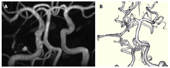
Figure 1 Three-dimensional time-of-flight magnetic resonance angiography.
A: Three-dimensional time-of-flight magnetic resonance angiography (3D-TOF-MRA) four years prior to admission; B: Volume rendering 3D-TOF-MRA study 1 year prior to admission, demonstrating two unruptured prenidal feeder artery aneurysms, one 8 mm × 3 mm fusiform proximal anterior inferior cerebral artery (AICA) aneurysm near the takeoff of the basilar artery and one 4.5 mm × 3 mm elliptical distal pedicle AICA aneurysm located at the entrance to the main inflow area of a small inconspicuous vascular nidus.
- Citation: Tucker A, Tsuji M, Yamada Y, Hanabusa K, Ukita T, Miyake H, Ohmura T. Arteriovenous malformation of the vestibulocochlear nerve. World J Clin Cases 2015; 3(7): 661-670
- URL: https://www.wjgnet.com/2307-8960/full/v3/i7/661.htm
- DOI: https://dx.doi.org/10.12998/wjcc.v3.i7.661


