Copyright
©The Author(s) 2015.
World J Clin Cases. Jul 16, 2015; 3(7): 556-574
Published online Jul 16, 2015. doi: 10.12998/wjcc.v3.i7.556
Published online Jul 16, 2015. doi: 10.12998/wjcc.v3.i7.556
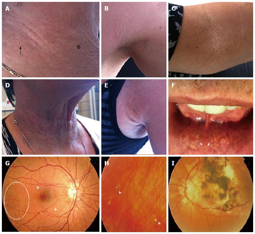
Figure 2 Dermatological (A-F) and ophthalmological (G-I) manifestations of pseudoxanthoma elasticum.
A, B: Flexural areas can show papular lesions (°) and coalesced plaques of papules (arrow); C: Cutaneous peau d’orange; D, E: Additional skin folds; F: Yellowish, reticular pattern on the mucosae of the lip (arrowed); G: Ocular fundi show peau d’orange (circle) and angioid streaks (arrowed); H: Comets and comet tails (arrowhead); I: Choroidal and subretinal hemorrhage.
- Citation: Vilder EYD, Vanakker OM. From variome to phenome: Pathogenesis, diagnosis and management of ectopic mineralization disorders. World J Clin Cases 2015; 3(7): 556-574
- URL: https://www.wjgnet.com/2307-8960/full/v3/i7/556.htm
- DOI: https://dx.doi.org/10.12998/wjcc.v3.i7.556


