Copyright
©The Author(s) 2015.
World J Clin Cases. Jun 16, 2015; 3(6): 519-524
Published online Jun 16, 2015. doi: 10.12998/wjcc.v3.i6.519
Published online Jun 16, 2015. doi: 10.12998/wjcc.v3.i6.519
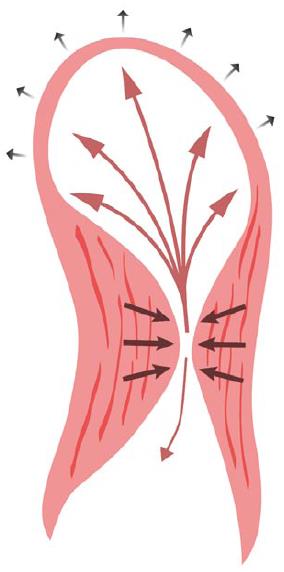
Figure 7 Illustration of blood flow direction during left ventricular systole.
While some blood is ejected towards the outflow tract (single arrow), the flow is initially greater towards the apex, which is the area of least resistance (arrows from mid-cavity to the left-ventricular apical aneurysm), resulting in a lower gradient across the mid-cavitary obliteration as the aneurysm size increases.
- Citation: Poulin MF, Shah A, Trohman RG, Madias C. Advanced Anderson-Fabry disease presenting with left ventricular apical aneurysm and ventricular tachycardia. World J Clin Cases 2015; 3(6): 519-524
- URL: https://www.wjgnet.com/2307-8960/full/v3/i6/519.htm
- DOI: https://dx.doi.org/10.12998/wjcc.v3.i6.519


