Copyright
©The Author(s) 2015.
World J Clin Cases. Jun 16, 2015; 3(6): 519-524
Published online Jun 16, 2015. doi: 10.12998/wjcc.v3.i6.519
Published online Jun 16, 2015. doi: 10.12998/wjcc.v3.i6.519
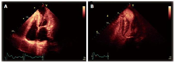
Figure 3 Transthoracic echocardiogram.
A: Transthoracic echocardiogram (TTE), apical four-chamber view, demonstrating concentric left ventricular hypertrophy with mid-cavitary obliteration at end-systole, as well as a large left-ventricular apical aneurysm (LVAA); B: TTE apical two-chamber view, with ultrasound contrast demonstrating stasis within the LVAA with a circular flow pattern, as well as decreased perfusion of the apex.
- Citation: Poulin MF, Shah A, Trohman RG, Madias C. Advanced Anderson-Fabry disease presenting with left ventricular apical aneurysm and ventricular tachycardia. World J Clin Cases 2015; 3(6): 519-524
- URL: https://www.wjgnet.com/2307-8960/full/v3/i6/519.htm
- DOI: https://dx.doi.org/10.12998/wjcc.v3.i6.519


