Copyright
©The Author(s) 2015.
World J Clin Cases. May 16, 2015; 3(5): 442-449
Published online May 16, 2015. doi: 10.12998/wjcc.v3.i5.442
Published online May 16, 2015. doi: 10.12998/wjcc.v3.i5.442
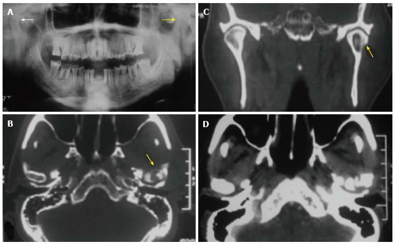
Figure 7 Chronic osteomyelitis of temporomandibular joint in a 55-year-old patient.
Sclerosis of left mandibular condyle is seen in panorex radiograph (A); a bony sequestrum is seen within the left mandibular condyle in axial (B) and coronal (C) reformatted computed tomography (CT) images; axial CT section in soft tissue window (D) shows inflammatory changes in tissues around the temporomandibular joint.
- Citation: Pahwa S, Bhalla AS, Roychaudhary A, Bhutia O. Multidetector computed tomography of temporomandibular joint: A road less travelled. World J Clin Cases 2015; 3(5): 442-449
- URL: https://www.wjgnet.com/2307-8960/full/v3/i5/442.htm
- DOI: https://dx.doi.org/10.12998/wjcc.v3.i5.442


