Copyright
©The Author(s) 2015.
World J Clin Cases. May 16, 2015; 3(5): 442-449
Published online May 16, 2015. doi: 10.12998/wjcc.v3.i5.442
Published online May 16, 2015. doi: 10.12998/wjcc.v3.i5.442
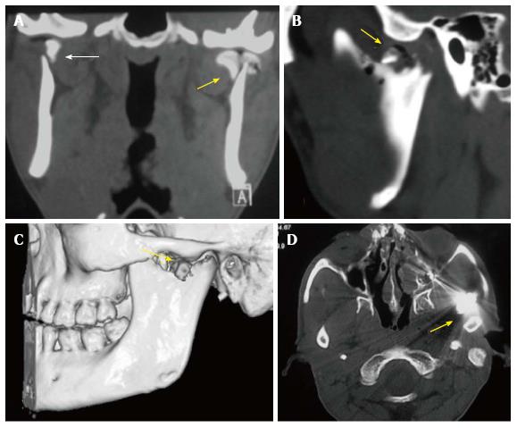
Figure 5 Condylar fractures in different patients.
Coronal (A), sagittal (B) and 3D (C) reconstructed CT images depict bilateral displaced, extracapsular (right- bold arrow), and intracapsular (left- arrow) condylar head fractures with involvement of the articular surface on the left side. The intra-articular air on the left side indicates an open wound (arrow). Open joint wound with a foreign body (bullet in another patient- D) or gross contamination are indications for open surgery.
- Citation: Pahwa S, Bhalla AS, Roychaudhary A, Bhutia O. Multidetector computed tomography of temporomandibular joint: A road less travelled. World J Clin Cases 2015; 3(5): 442-449
- URL: https://www.wjgnet.com/2307-8960/full/v3/i5/442.htm
- DOI: https://dx.doi.org/10.12998/wjcc.v3.i5.442


