Copyright
©The Author(s) 2015.
World J Clin Cases. May 16, 2015; 3(5): 405-417
Published online May 16, 2015. doi: 10.12998/wjcc.v3.i5.405
Published online May 16, 2015. doi: 10.12998/wjcc.v3.i5.405
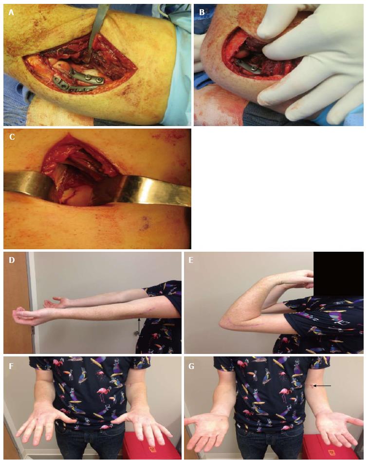
Figure 5 Two-incision approach.
A case demonstrating the two incision approach (lateral and medial). A: The lateral collateral ligament is not reflected (the plate is placed directly on top of it). Stable fixation of the capitellar fragment could not be obtained with headless compression screws and a one-third tubular plate was added for stability; B: Flexion demonstrates a mild amount of impingement, which was not symptomatic postoperatively; C: Anterior view demonstrates trochlear fracture line. Even though two incisions are used there is less soft tissue dissection overall than required in other approaches; D-G: Approximate 6 wk follow-up range of motion in this patient.
- Citation: Yari SS, Bowers NL, Craig MA, Reichel LM. Management of distal humeral coronal shear fractures. World J Clin Cases 2015; 3(5): 405-417
- URL: https://www.wjgnet.com/2307-8960/full/v3/i5/405.htm
- DOI: https://dx.doi.org/10.12998/wjcc.v3.i5.405


