Copyright
©The Author(s) 2015.
World J Clin Cases. Apr 16, 2015; 3(4): 389-392
Published online Apr 16, 2015. doi: 10.12998/wjcc.v3.i4.389
Published online Apr 16, 2015. doi: 10.12998/wjcc.v3.i4.389
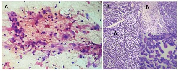
Figure 2 Photomicrograph.
A: Photomicrograph of cytological smear showing spindle cells with pleomorphic hyperchromatic nuclei and a plump cell (arrow) with abundant eosinophilic cytoplasm; B: Photomicrograph showing hypercellular areas (Antoni A) of spindle cells having hyperchromatic nuclei and hypocellular areas (Antoni B) (100 ×). Inset shows rhabdomyoblastic differentiation (400 ×).
- Citation: Shete S, Bolde S, Pandit G, Matkari P, Ingle SB. Unusual histological variant of malignant peripheral nerve sheath tumor with rhabdomyoblastic differentiation. World J Clin Cases 2015; 3(4): 389-392
- URL: https://www.wjgnet.com/2307-8960/full/v3/i4/389.htm
- DOI: https://dx.doi.org/10.12998/wjcc.v3.i4.389


