Copyright
©The Author(s) 2015.
World J Clin Cases. Mar 16, 2015; 3(3): 231-244
Published online Mar 16, 2015. doi: 10.12998/wjcc.v3.i3.231
Published online Mar 16, 2015. doi: 10.12998/wjcc.v3.i3.231
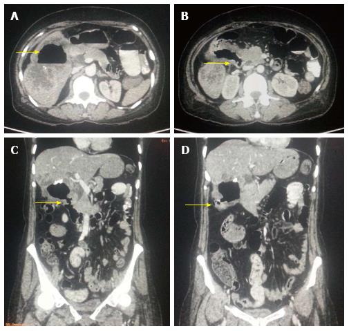
Figure 2 Aggressive gallbladder cancer and entero-biliary fistula.
A: Axial contrast-enhanced computed tomography (CECT) abdominal section shows predominantly hypodense mass lesion replacing gall bladder fossa with presence of air (arrow) raising suspicion of fistula formation. Adjacent hepatic segments are also infiltrated by the mass; B and C: Axial and Coronal CECT abdominal sections clearly reveal the fistulous communication with D2 segment of duodenum (arrow); D: Coronal CECT abdominal section also show coexisting cholecysto-colonic fistula with hepatic flexure of colon (arrow).
- Citation: Dwivedi AND, Jain S, Dixit R. Gall bladder carcinoma: Aggressive malignancy with protean loco-regional and distant spread. World J Clin Cases 2015; 3(3): 231-244
- URL: https://www.wjgnet.com/2307-8960/full/v3/i3/231.htm
- DOI: https://dx.doi.org/10.12998/wjcc.v3.i3.231


