Copyright
©The Author(s) 2015.
World J Clin Cases. Feb 16, 2015; 3(2): 191-195
Published online Feb 16, 2015. doi: 10.12998/wjcc.v3.i2.191
Published online Feb 16, 2015. doi: 10.12998/wjcc.v3.i2.191
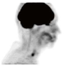
Figure 4 Positron emission tomography performed at four months post-treatment.
The Positron emission tomography shows no definite or abnormal fluorodeoxyglucose (FDG) activity to suggest the presence of metabolically active tumor with special attention to the ethmoidal region adjacent to the cribriform plate. Linear FDG activity in the distal esophagus likely represents esophagitis.
- Citation: Noticewala SS, Mell LK, Olson SE, Read W. Survival in unresectable sinonasal undifferentiated carcinoma treated with concurrent intra-arterial cisplatin and radiation. World J Clin Cases 2015; 3(2): 191-195
- URL: https://www.wjgnet.com/2307-8960/full/v3/i2/191.htm
- DOI: https://dx.doi.org/10.12998/wjcc.v3.i2.191


