Copyright
©The Author(s) 2015.
World J Clin Cases. Feb 16, 2015; 3(2): 191-195
Published online Feb 16, 2015. doi: 10.12998/wjcc.v3.i2.191
Published online Feb 16, 2015. doi: 10.12998/wjcc.v3.i2.191
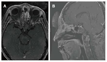
Figure 3 T1 Magnetic resonance imaging after completing fourth dose of chemotherapy.
A: Axial T1 magnetic resonance imaging (MRI) and B: Sagittal T1 MRI show the mass had decreased in size as compared to the prior to treatment MRI. The mediolateral dimension is 2.23 cm which is decreased in size from the prior examination at which time it measured 3.32 cm. The AP appears to have decreased in size to 2.42 cm as compared to 4.93 cm on the prior MRI. There appears to be residual enhancing tissue in the right posterior ethmoid. The intracranial enhancement and edema within the inferior left frontal lobe is significantly decreased.
- Citation: Noticewala SS, Mell LK, Olson SE, Read W. Survival in unresectable sinonasal undifferentiated carcinoma treated with concurrent intra-arterial cisplatin and radiation. World J Clin Cases 2015; 3(2): 191-195
- URL: https://www.wjgnet.com/2307-8960/full/v3/i2/191.htm
- DOI: https://dx.doi.org/10.12998/wjcc.v3.i2.191


