Copyright
©The Author(s) 2015.
World J Clin Cases. Nov 16, 2015; 3(11): 956-964
Published online Nov 16, 2015. doi: 10.12998/wjcc.v3.i11.956
Published online Nov 16, 2015. doi: 10.12998/wjcc.v3.i11.956
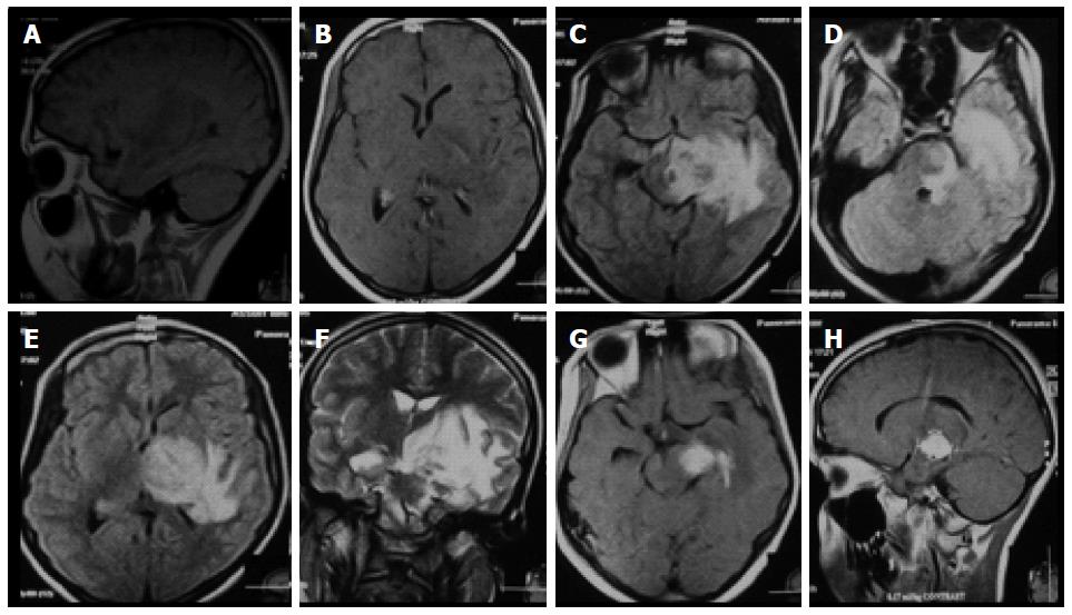
Figure 1 Cranial magnetic resonance imaging brain (on admission at September 2009) showing (A, B) sagittal and axial T1-weighted views with a solitary hypointense lesion in the left temporal lobe; (C-E) axial fluid attenuated inversion recovery and T2-weighted (F) images showing hyperintense lesion in the left temporal lobe encroaching on the adjacent brainstem with perifocal edema and mass effect; (G, H) axial and sagittal T1-weighted views showing homogenous solitary enhanced lesion in the left temporal lobe encroaching on the adjacent brainstem and surrounded by a moderate hypointensity.
- Citation: Hamed SA, Mekkawy MA, Abozaid H. Differential diagnosis of a vanishing brain space occupying lesion in a child. World J Clin Cases 2015; 3(11): 956-964
- URL: https://www.wjgnet.com/2307-8960/full/v3/i11/956.htm
- DOI: https://dx.doi.org/10.12998/wjcc.v3.i11.956


