Copyright
©2014 Baishideng Publishing Group Inc.
World J Clin Cases. Aug 16, 2014; 2(8): 385-390
Published online Aug 16, 2014. doi: 10.12998/wjcc.v2.i8.385
Published online Aug 16, 2014. doi: 10.12998/wjcc.v2.i8.385
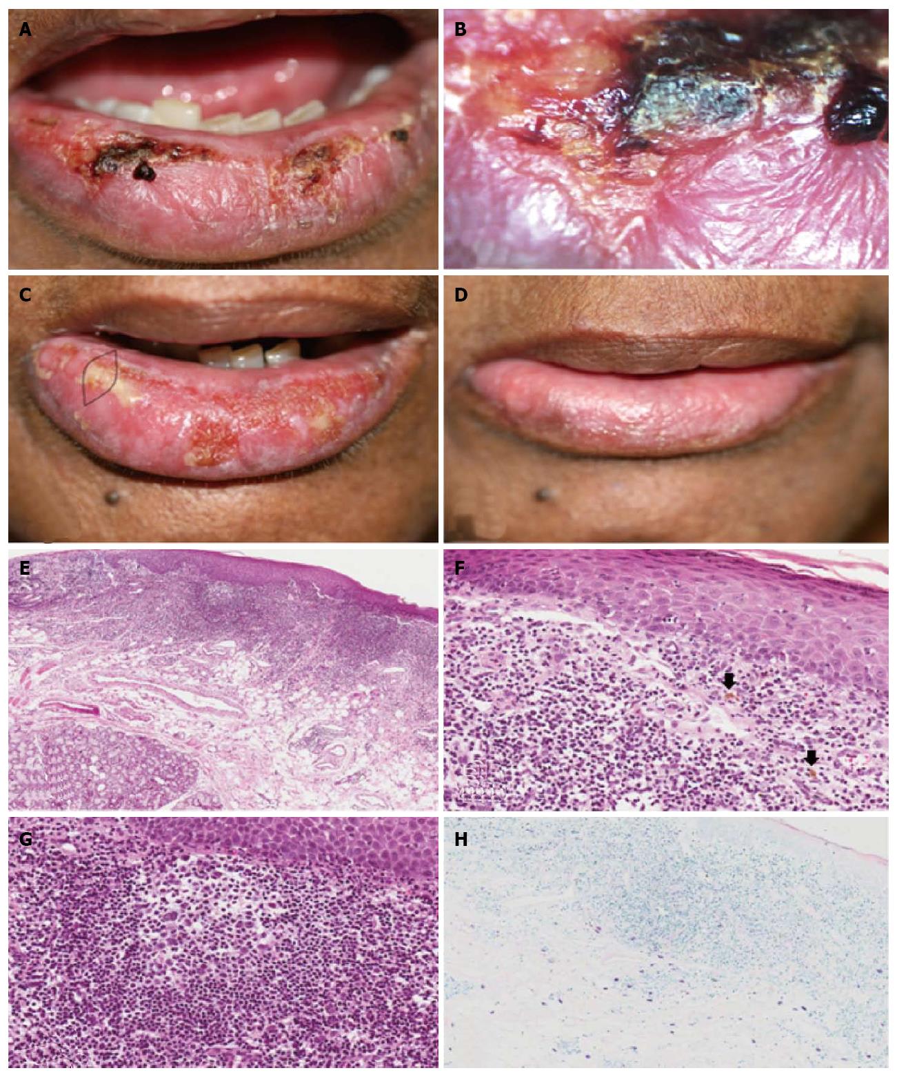
Figure 1 Case 1.
A: Clinical aspect at the first appointment, showing lower lip edema, ulcers and crusts; B: Videoroscopy image showing in detail the presence of ulcer and crust; C: Clinical aspect at the second appointment showing the area of biopsy; D: Clinical aspect one month after treatment, showing remission of the lip edema, ulcers and crusts; E: Histological aspects. Epithelial atrophy and intense diffuse lymphoplasmacytic inflammatory infiltrate extending deep into the fatty tissue (× 10, HE); F: Epithelium showing spongiosis, hydropic degeneration of the basal layer cells and lymphocytic exocytosis. In the connective tissue, lymphocytic inflammatory infiltrate and pigmentary incontinence (arrows) were observed (× 40, HE). G: Secondary lymphoid follicle (× 40, HE); H: Mast cells mainly in the deeper area of the connective tissue (× 20, Giemsa).
- Citation: Miranda AM, Ferrari TM, Werneck JT, Junior AS, Cunha KS, Dias EP. Actinic prurigo of the lip: Two case reports. World J Clin Cases 2014; 2(8): 385-390
- URL: https://www.wjgnet.com/2307-8960/full/v2/i8/385.htm
- DOI: https://dx.doi.org/10.12998/wjcc.v2.i8.385


