Copyright
©2014 Baishideng Publishing Group Inc.
World J Clin Cases. Aug 16, 2014; 2(8): 380-384
Published online Aug 16, 2014. doi: 10.12998/wjcc.v2.i8.380
Published online Aug 16, 2014. doi: 10.12998/wjcc.v2.i8.380
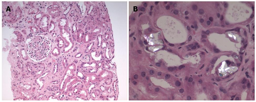
Figure 2 Hematoxylin and eosin stain.
A: Hematoxylin and eosin stain demonstrating moderate interstitial fibrosis and tubular atrophy; B: Hematoxylin and eosin stain with polarized light demonstrating calcium oxalate deposition within the tubules.
- Citation: Sivabalasundaram V, Habal F, Cherney D. Prucalopride-associated acute tubular necrosis. World J Clin Cases 2014; 2(8): 380-384
- URL: https://www.wjgnet.com/2307-8960/full/v2/i8/380.htm
- DOI: https://dx.doi.org/10.12998/wjcc.v2.i8.380


