Copyright
©2014 Baishideng Publishing Group Inc.
World J Clin Cases. Aug 16, 2014; 2(8): 367-372
Published online Aug 16, 2014. doi: 10.12998/wjcc.v2.i8.367
Published online Aug 16, 2014. doi: 10.12998/wjcc.v2.i8.367
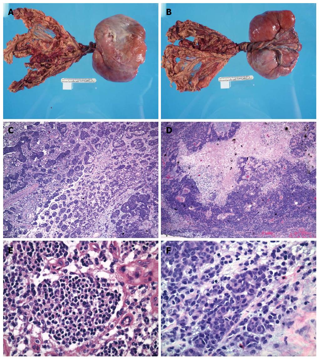
Figure 1 Gross and microscopic feature of abdominal mass.
A, B: Gross examination showed an encapsulated and lobulated soft tissue mass with attached omental tissue; C-F: The small round cell tumor possessed round to oval hyperchromatic nuclei with inconspicuous nucleoli and scant cytoplasm. Focal tumor cells with enlarged nuclei, open chromatin and prominent nucleoli, as well as rhabdoid differentiation were noted. Focal tumor necrosis and hemorrhages were present (HE staining).
- Citation: Liang L, Tatevian N, Bhattacharjee M, Tsao K, Hicks J. Desmoplastic small round cell tumor with atypical immunohistochemical profile and rhabdoid-like differentiation. World J Clin Cases 2014; 2(8): 367-372
- URL: https://www.wjgnet.com/2307-8960/full/v2/i8/367.htm
- DOI: https://dx.doi.org/10.12998/wjcc.v2.i8.367


