Copyright
©2014 Baishideng Publishing Group Inc.
World J Clin Cases. Jun 16, 2014; 2(6): 219-223
Published online Jun 16, 2014. doi: 10.12998/wjcc.v2.i6.219
Published online Jun 16, 2014. doi: 10.12998/wjcc.v2.i6.219
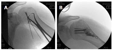
Figure 4 Intra-operative fluoroscopic images.
Initially, the bigger fragments of the lesser tuberosity were reduced using reduction forceps and fixed using cannulated headless Hebert screws (A). The smaller fragments were then reduced and stabilized under a low profile, bendable neutralization buttress plate (B).
- Citation: Nikolaou VS, Chytas D, Tyrpenou E, Babis GC. Two-level reconstruction of isolated fracture of the lesser tuberosity of the humerus. World J Clin Cases 2014; 2(6): 219-223
- URL: https://www.wjgnet.com/2307-8960/full/v2/i6/219.htm
- DOI: https://dx.doi.org/10.12998/wjcc.v2.i6.219


