Copyright
©2014 Baishideng Publishing Group Inc.
World J Clin Cases. Jun 16, 2014; 2(6): 206-208
Published online Jun 16, 2014. doi: 10.12998/wjcc.v2.i6.206
Published online Jun 16, 2014. doi: 10.12998/wjcc.v2.i6.206
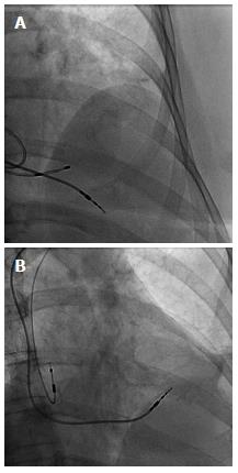
Figure 4 Image.
Top image shows the right ventricular permanent pacemaker lead protruding well past the heart border, A temporary transvenous pacemaker lead can also be seen within the right ventricle (A); The bottom image shows the repositioned right ventricular lead higher up on the interventricular septum and absence of the temporary transvenous pacemaker lead. The right atrial lead can also be seen in this image (B).
- Citation: Nash G, Williams JM, Nekkanti R, Movahed A. Case of early right ventricular pacing lead perforation and review of the literature. World J Clin Cases 2014; 2(6): 206-208
- URL: https://www.wjgnet.com/2307-8960/full/v2/i6/206.htm
- DOI: https://dx.doi.org/10.12998/wjcc.v2.i6.206


