Copyright
©2014 Baishideng Publishing Group Inc.
World J Clin Cases. Jun 16, 2014; 2(6): 206-208
Published online Jun 16, 2014. doi: 10.12998/wjcc.v2.i6.206
Published online Jun 16, 2014. doi: 10.12998/wjcc.v2.i6.206
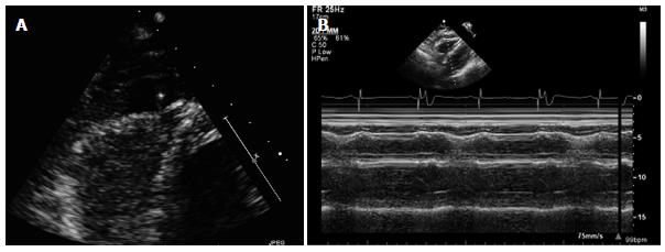
Figure 2 Transthoracic echocardiogram.
A: Image is an apical 4 chamber view of a transthoracic echocardiogram showing a moderate sized localized pericardial effusion with an echo bright structure within the effusion suspicious for lead perforation; B: Image is taken in M-mode and demonstrates right ventricular diastolic collapse suggestive of increased pericardial pressures.
- Citation: Nash G, Williams JM, Nekkanti R, Movahed A. Case of early right ventricular pacing lead perforation and review of the literature. World J Clin Cases 2014; 2(6): 206-208
- URL: https://www.wjgnet.com/2307-8960/full/v2/i6/206.htm
- DOI: https://dx.doi.org/10.12998/wjcc.v2.i6.206


