Copyright
©2014 Baishideng Publishing Group Inc.
World J Clin Cases. May 16, 2014; 2(5): 146-150
Published online May 16, 2014. doi: 10.12998/wjcc.v2.i5.146
Published online May 16, 2014. doi: 10.12998/wjcc.v2.i5.146
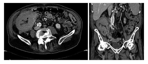
Figure 2 Computed tomography exam in the axial and coronal planes.
The images show the concentric thickening of the wall of the cecum and ascending colon. The millimetric air bubbles in the context of the colic wall are also depicted.
- Citation: Gigli S, Buonocore V, Barchetti F, Glorioso M, Di Brino M, Guerrisi P, Buonocore C, Giovagnorio F, Giraldi G. Primary colonic lymphoma: An incidental finding in a patient with a gallstone attack. World J Clin Cases 2014; 2(5): 146-150
- URL: https://www.wjgnet.com/2307-8960/full/v2/i5/146.htm
- DOI: https://dx.doi.org/10.12998/wjcc.v2.i5.146


