Copyright
©2014 Baishideng Publishing Group Inc.
World J Clin Cases. May 16, 2014; 2(5): 126-132
Published online May 16, 2014. doi: 10.12998/wjcc.v2.i5.126
Published online May 16, 2014. doi: 10.12998/wjcc.v2.i5.126
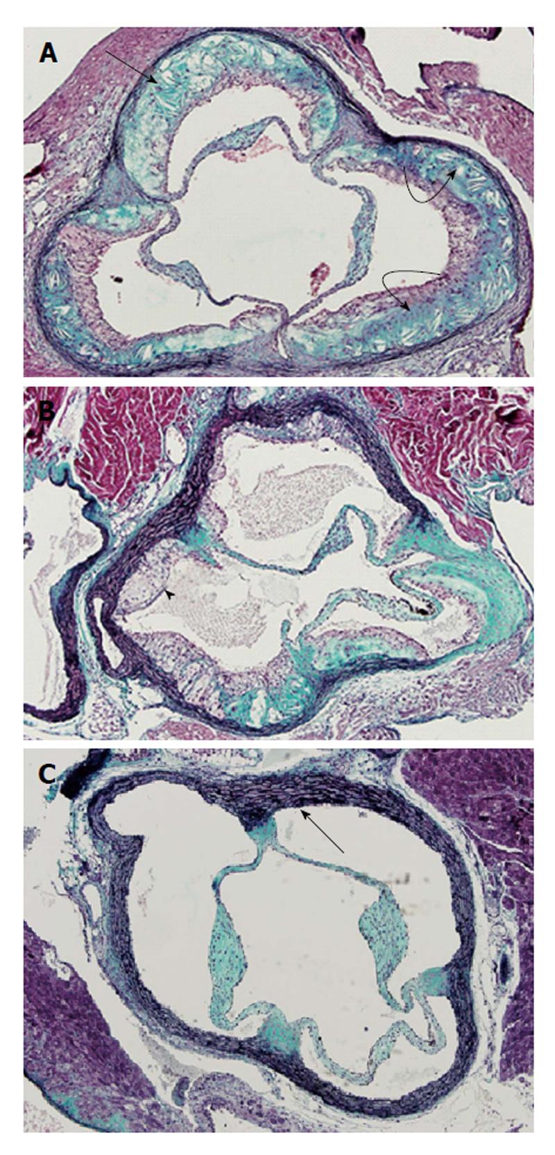
Figure 1 Representative photomicrographs taken at aortic root from apolipoprotein E-knockout (A), low density lipoprotein-r-knockout (B) and their wild-type background C57BL/6J (C) mice.
A: Advanced atherosclerosis lesions in all 3 valves of the aortic root (arrow); these lesions are composed of numerous cholesterol clefts (curved arrows); B: Illustrating lipid-rich atherosclerotic lesions in the aortic root; the arrow head points to an atherosclerotic plaque primarily composed of apparent foam cells. No cholesterol cleft is visible in B; C: Demonstrating an atherosclerotic-lesion-free aortic root with normal-looking vascular wall (arrow) with apparent intact elastic lamina and endothelium. Trichrome staining; × 40.
- Citation: Kapourchali FR, Surendiran G, Chen L, Uitz E, Bahadori B, Moghadasian MH. Animal models of atherosclerosis. World J Clin Cases 2014; 2(5): 126-132
- URL: https://www.wjgnet.com/2307-8960/full/v2/i5/126.htm
- DOI: https://dx.doi.org/10.12998/wjcc.v2.i5.126


