Copyright
©2014 Baishideng Publishing Group Co.
World J Clin Cases. Apr 16, 2014; 2(4): 111-119
Published online Apr 16, 2014. doi: 10.12998/wjcc.v2.i4.111
Published online Apr 16, 2014. doi: 10.12998/wjcc.v2.i4.111
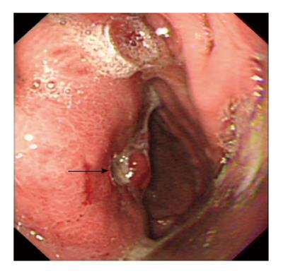
Figure 2 Gastroscopy revealing a protruding lesion on the back wall of the gastric cardia and fundus with a coarse surface, and an approximately 1.
0 cm × 1.0 cm ulcer was located in the middle of the lesion (black arrow).
- Citation: Yi XJ, Chen CQ, Li Y, Ma JP, Li ZX, Cai SR, He YL. Rare case of an abdominal mass: Reactive nodular fibrous pseudotumor of the stomach encroaching on multiple abdominal organs. World J Clin Cases 2014; 2(4): 111-119
- URL: https://www.wjgnet.com/2307-8960/full/v2/i4/111.htm
- DOI: https://dx.doi.org/10.12998/wjcc.v2.i4.111


