Copyright
©2014 Baishideng Publishing Group Inc.
World J Clin Cases. Nov 16, 2014; 2(11): 623-641
Published online Nov 16, 2014. doi: 10.12998/wjcc.v2.i11.623
Published online Nov 16, 2014. doi: 10.12998/wjcc.v2.i11.623
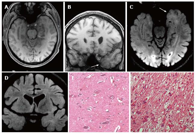
Figure 11 Gangliogliomas World Health Organization grade I of the left temporo-mesial region and focal cortical dysplasia IIa subtype associated.
Axial and coronal T1-w images (A-B) show thickening of amygdala and uncus (arrow in B). Axial and coronal FLAIR T2-w images (C-D) present blurring and adjacent subcortical high signal abnormality compatible with a focal cortical dysplasia (FCD). Microscopy evidenced a glioneuronal tumor, with scattered binucleated ganglion cells, compatible with a gan-glioglioma (E) and dysmorphic neurons in the adjacent cortex (FCD Type IIa) (F).
- Citation: Giulioni M, Marucci G, Martinoni M, Marliani AF, Toni F, Bartiromo F, Volpi L, Riguzzi P, Bisulli F, Naldi I, Michelucci R, Baruzzi A, Tinuper P, Rubboli G. Epilepsy associated tumors: Review article. World J Clin Cases 2014; 2(11): 623-641
- URL: https://www.wjgnet.com/2307-8960/full/v2/i11/623.htm
- DOI: https://dx.doi.org/10.12998/wjcc.v2.i11.623


