Copyright
©2014 Baishideng Publishing Group Inc.
World J Clin Cases. Nov 16, 2014; 2(11): 623-641
Published online Nov 16, 2014. doi: 10.12998/wjcc.v2.i11.623
Published online Nov 16, 2014. doi: 10.12998/wjcc.v2.i11.623
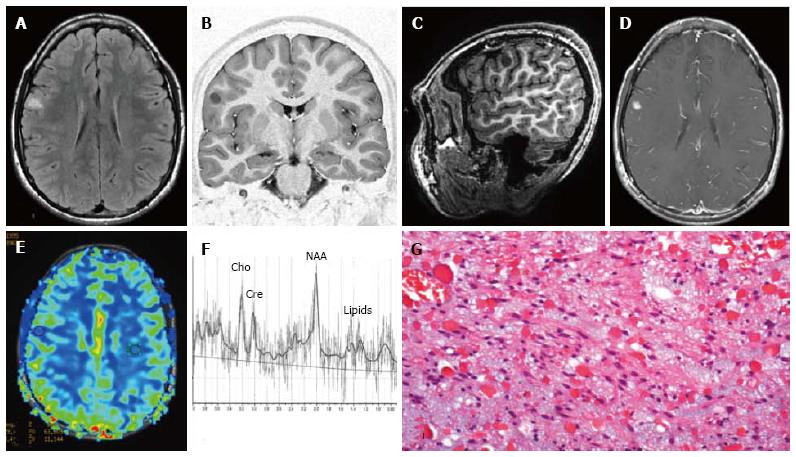
Figure 8 Pilocytic astrocytoma World Health Organization grade I of the right frontal lobe.
Axial FLAIR T2-w (A) and coronal lR T1-w (B) images show a cortical-subcortical lesion, with cystic component, with minimal mass effect. The tumor appears well de-marcated (C) on 3D sagittal sequence and displays nodular and homogeneous en-hancement on post-contrast axial T1-w images (D). Perfusion study doesn’t show any rCBV increase within the lesion (E). MR Spectroscopy (MRS) study (F) reveals elevation in choline and reduction in NAA. (G) Histological examination shows a tumor com-posed of areas rich in myxoid material, elongated glial elements with uniform nuclei and numerous eosinophilic granular bodies.
- Citation: Giulioni M, Marucci G, Martinoni M, Marliani AF, Toni F, Bartiromo F, Volpi L, Riguzzi P, Bisulli F, Naldi I, Michelucci R, Baruzzi A, Tinuper P, Rubboli G. Epilepsy associated tumors: Review article. World J Clin Cases 2014; 2(11): 623-641
- URL: https://www.wjgnet.com/2307-8960/full/v2/i11/623.htm
- DOI: https://dx.doi.org/10.12998/wjcc.v2.i11.623


