Copyright
©2014 Baishideng Publishing Group Inc.
World J Clin Cases. Nov 16, 2014; 2(11): 623-641
Published online Nov 16, 2014. doi: 10.12998/wjcc.v2.i11.623
Published online Nov 16, 2014. doi: 10.12998/wjcc.v2.i11.623
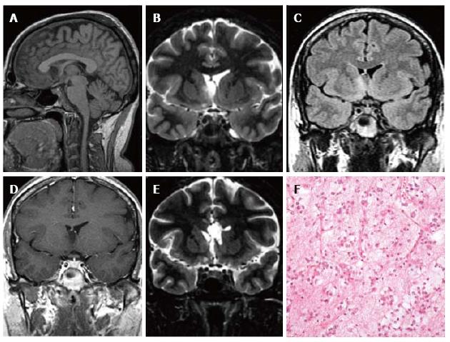
Figure 6 Extra temporal DNET.
Sagittal 3D T1 (A) coronal IR T1 (B) and FLAIR T2-w (C) demonstrate a cystic wedge-shaped lesion in the right fronto-orbital gyrus. On FLAIR T2-w the tumor is slightly hypointense with a faint hyperintense rim. On post contrast coronal T1-w images there is no enhancement uptake (D); E: Post-surgical scan on cor-onal IR T1-w; F: Microscopic study evidences the presence of floating neurons, a feature of DNET, in microcystic areas lined with oligo-like cells.
- Citation: Giulioni M, Marucci G, Martinoni M, Marliani AF, Toni F, Bartiromo F, Volpi L, Riguzzi P, Bisulli F, Naldi I, Michelucci R, Baruzzi A, Tinuper P, Rubboli G. Epilepsy associated tumors: Review article. World J Clin Cases 2014; 2(11): 623-641
- URL: https://www.wjgnet.com/2307-8960/full/v2/i11/623.htm
- DOI: https://dx.doi.org/10.12998/wjcc.v2.i11.623


