Copyright
©2014 Baishideng Publishing Group Inc.
World J Clin Cases. Nov 16, 2014; 2(11): 623-641
Published online Nov 16, 2014. doi: 10.12998/wjcc.v2.i11.623
Published online Nov 16, 2014. doi: 10.12998/wjcc.v2.i11.623
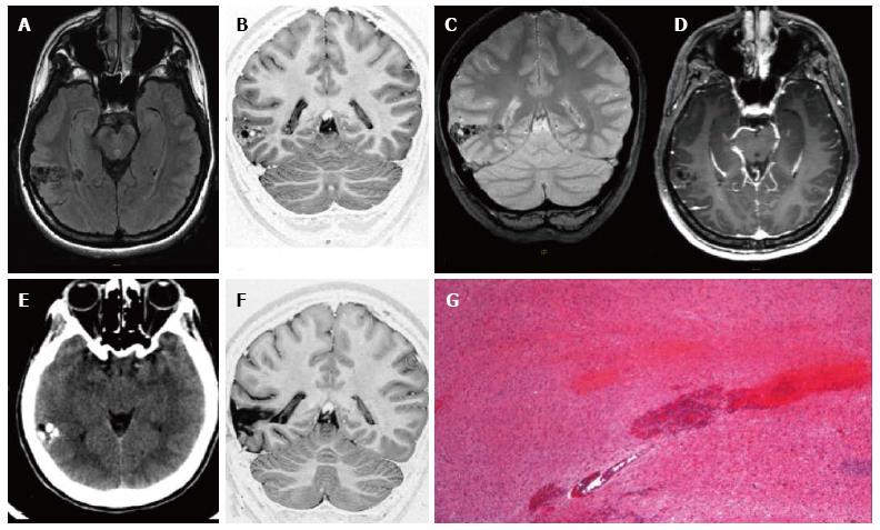
Figure 1 Ganglioglioma World Health Organization grade I of the right posterior middle temporal gyrus.
Axial FLAIR T2-w (A) and coronal IR T1-w (B) images show inhomogeneous cortical-subcortical mass extending within the deep white matter and reaching the ependymal layer. The tumor presents a combination of solid, cystic and calcified components. The latter is better identified on coronal T2*-w sequence (C). Post-contrast axial T1-w image (D) shows no pathological enhancement and axial CT scan (E) confirms the calcified component. Coronal IR T1-w image (F) demonstrates lesion resec-tion; G: Histological examination evidences a biphasic neoplastic population, with neu-ronal and glial elements.
- Citation: Giulioni M, Marucci G, Martinoni M, Marliani AF, Toni F, Bartiromo F, Volpi L, Riguzzi P, Bisulli F, Naldi I, Michelucci R, Baruzzi A, Tinuper P, Rubboli G. Epilepsy associated tumors: Review article. World J Clin Cases 2014; 2(11): 623-641
- URL: https://www.wjgnet.com/2307-8960/full/v2/i11/623.htm
- DOI: https://dx.doi.org/10.12998/wjcc.v2.i11.623


